Education and Training


Photoacoustic Imaging in Detecting Ovarian or Fallopian Tube Cancer
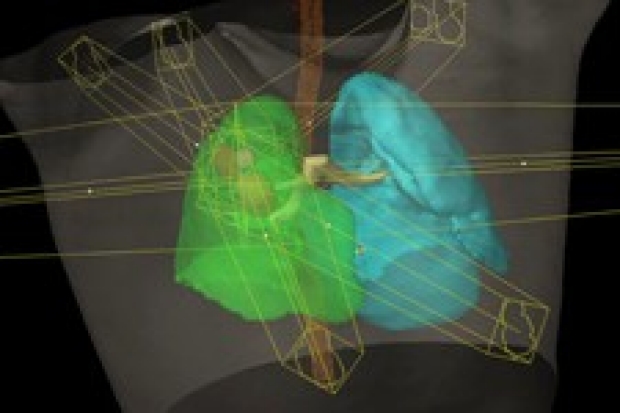
This pilot clinical trial studies how well photoacoustic imaging works in detecting ovarian or fallopian tube cancer. Photoacoustic imaging is an imaging method that uses lasers to light up tissue, and then converts the light information into ultrasound images. Photoacoustic imaging can provide images of the structure of tissues, as well as their function and the levels of molecules, such as the flow of blood in blood vessels and the level of oxygen in the blood. Photoacoustic imaging may help doctors determine whether a mass is benign (non-cancerous) or cancerous based on the molecular differences between cancer and normal tissue. It may be more accurate and less expensive than other imaging methods, and does not expose patients to radiation.
Stanford is currently not accepting patients for this trial.
Stanford Investigator(s):
Intervention(s):
- procedure: Photoacoustic Imaging
Eligibility
Inclusion Criteria:
- Patients must be undergoing ovarian resection
- Ability to understand and the willingness to sign a written informed consent document
Exclusion Criteria:
- Patients who have had primary surgical excision
- Pregnant or lactating women
Ages Eligible for Study
18 Years - 80 Years
Genders Eligible for Study
Female
Not currently accepting new patients for this trial
Contact Information
Stanford University
School of Medicine
300 Pasteur Drive
Stanford,
CA
94305
Sri-Rajasekhar Kothapalli
Not Recruiting